1
10–20 years before symptoms
For Clinicians
White Scan turns a structural brain MRI into Alzheimer's pathology insight—with an automated amyloid report.
Book a DemoThe Clinical Gap
10–20 years before symptoms
Pre-symptomatic
Symptomatic
Clinical pathway
Where White Scan adds clinical value
White Scan uses are subject to ongoing clinical validation and applicable regulatory requirements.
White Scan supports assessment before and after symptom presentation. It does not provide a standalone diagnosis.
Beyond Routine MRI
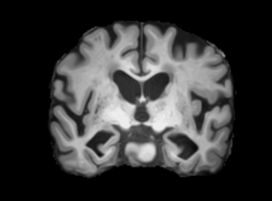
Shows brain anatomy and structural change for radiologist interpretation.
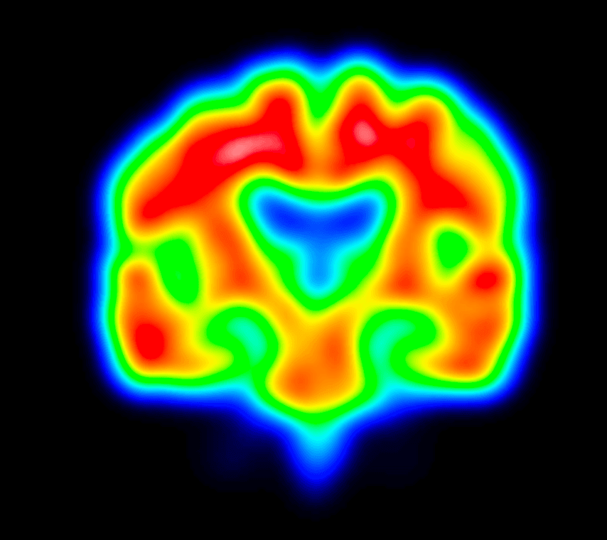
Adds MRI-derived surrogate amyloid burden and an automatically generated report.
Two Clinical Uses
For appropriate asymptomatic or at-risk adults as part of clinician-led assessment.
Supports differential diagnosis, clinical phenotyping and treatment planning within the full clinical context.
The Analysis Report
An explicit amyloid-positive or amyloid-negative result.
A composite score with threshold-based classification.
Indications of regional amyloid deposition across the brain.
The fully automated White Scan report must be interpreted alongside clinical assessment, history, the radiologist's MRI report, volumetry and other relevant information.
Imaging Input
Evaluated against gold-standard biomarkers
Evaluated against established biomarker references.
Amyloid PETAV45 florbetapir
Cerebrospinal fluidtTau/Aβ42
Validation studies are in progress at the Brain Health Clinic at South London and Maudsley NHS Foundation Trust to support wider clinical adoption.
Research and clinical partners




See White Scan in practice
Online demos can be booked directly. In-person demos are subject to availability and location—please contact us.
Book a DemoAlready Have an MRI?
For a complete whole-brain 3D T1-weighted MRI in DICOM format, contact us for secure transfer instructions.
Email Silver Matter